MicroCT scan of the skull of an Asian elephant (Elephas maximus). Dataset to Grunstra et al. (2024) "Convergent evolution in Afrotheria and non-afrotherians demonstrates high evolvability of the mammalian inner ear"
MicroCT scan of the skull of an Asian elephant (Elephas maximus). This dataset was part of a study on the evolution of the inner ear.
Usage Notes
The skull was scanned in two parts. "skull 1" covers the area of the inner ear that was studied in the related publication by Grunstra et al. (2024).
X-ray tube: FXE Direct Beam
Detector: Perkin Elmer Y.Panel 4343 CT
Exposure time: 1 s for 2430 projections per scan
Voltage: 135 kV
Current: 110 µA
Filter: 1 mm Copper
Reconstructed voxel size: 95.6 µm
The image stack was cropped and converted into a TIF image stack using the software Dragonfly.
This specimen was used in a study on the inner ear (see "Publications") the segmented inner ear data is available here: https://osf.io/9mtwh/
Funding
Agency
Program
Proj. Id
Proj. Title
FWF Austrian Science Fund
P 33736
Evolvabilität des Innen- & Mittelohrs in Vögeln und Säugern
Publications
Please cite the following publication when reusing this dataset:
Grunstra, N.D.S., Hollinetz, F., Bravo Morante, G. et al. Convergent evolution in Afrotheria and non-afrotherians demonstrates high evolvability of the mammalian inner ear.
Nat Commun15, 7869 (2024). https://doi.org/10.1038/s41467-024-52180-1
Grunstra,
Nicole D, Fabian Hollinetz, Guillermo B Morante, Frank Zachos, Cathrin
Pfaff, Viola Winkler, Philipp Mitteroecker, and Anne Le Maître. 2024.
“Data for ‘Convergent Evolution in Afrotheria and Non-Afrotherians
Demonstrates High Evolvability of the Mammalian Inner Ear.’” OSF. August
28. osf.io/9mtwh.
TIF image stack of the microCT scan of the second part ("skull 2") of the Asian elephant skull
15822 MB
Media
Images
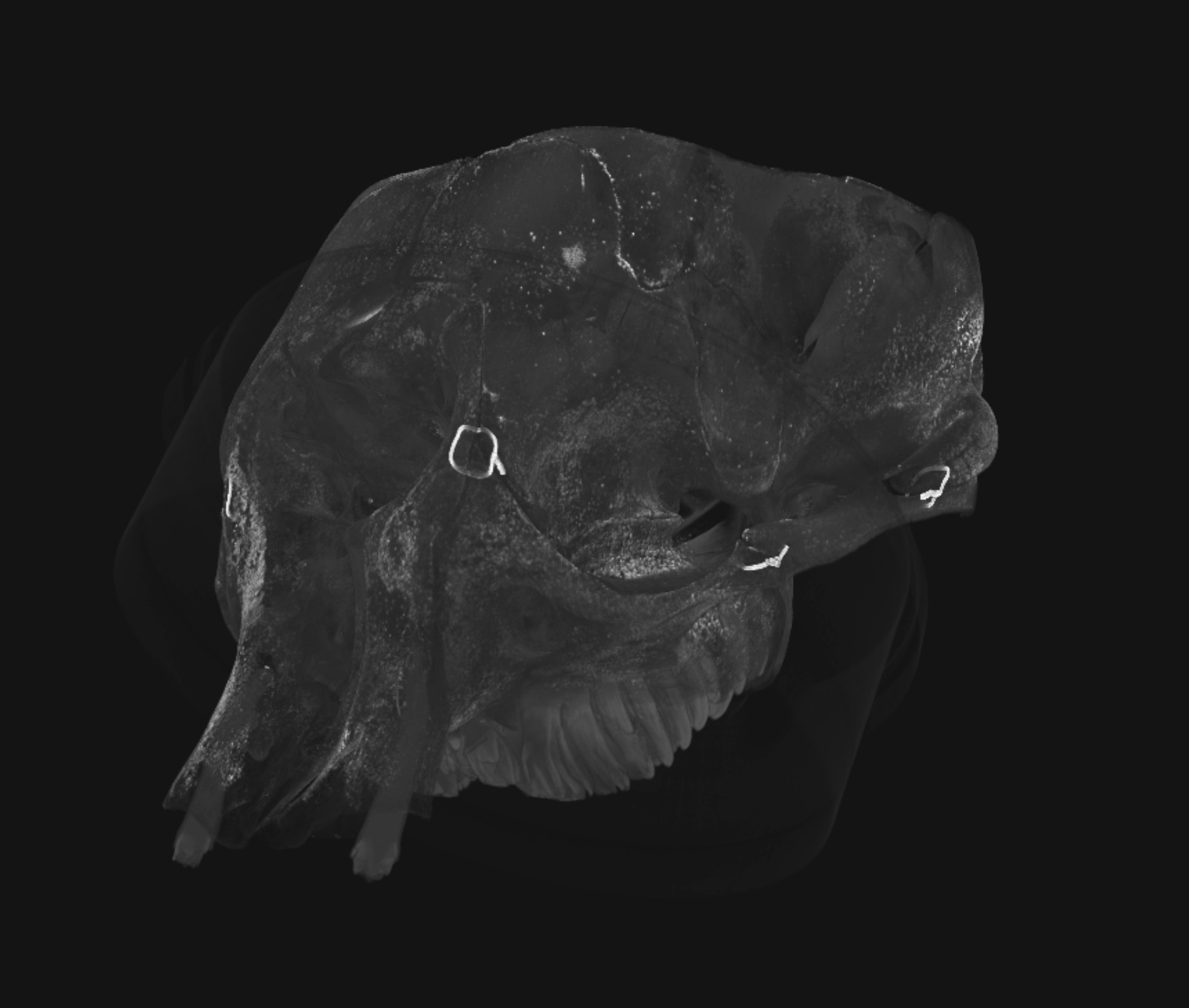
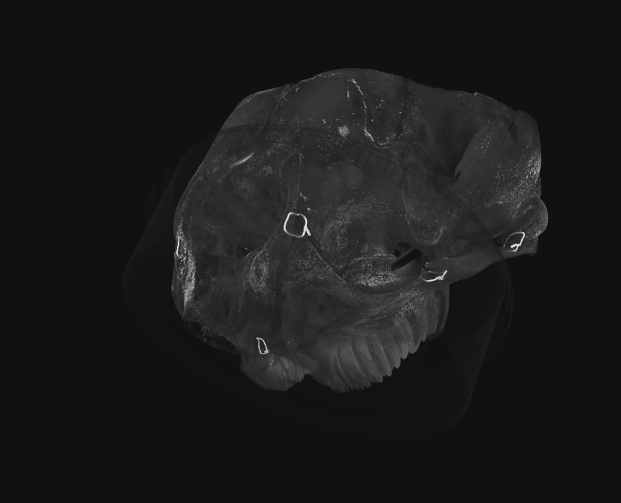
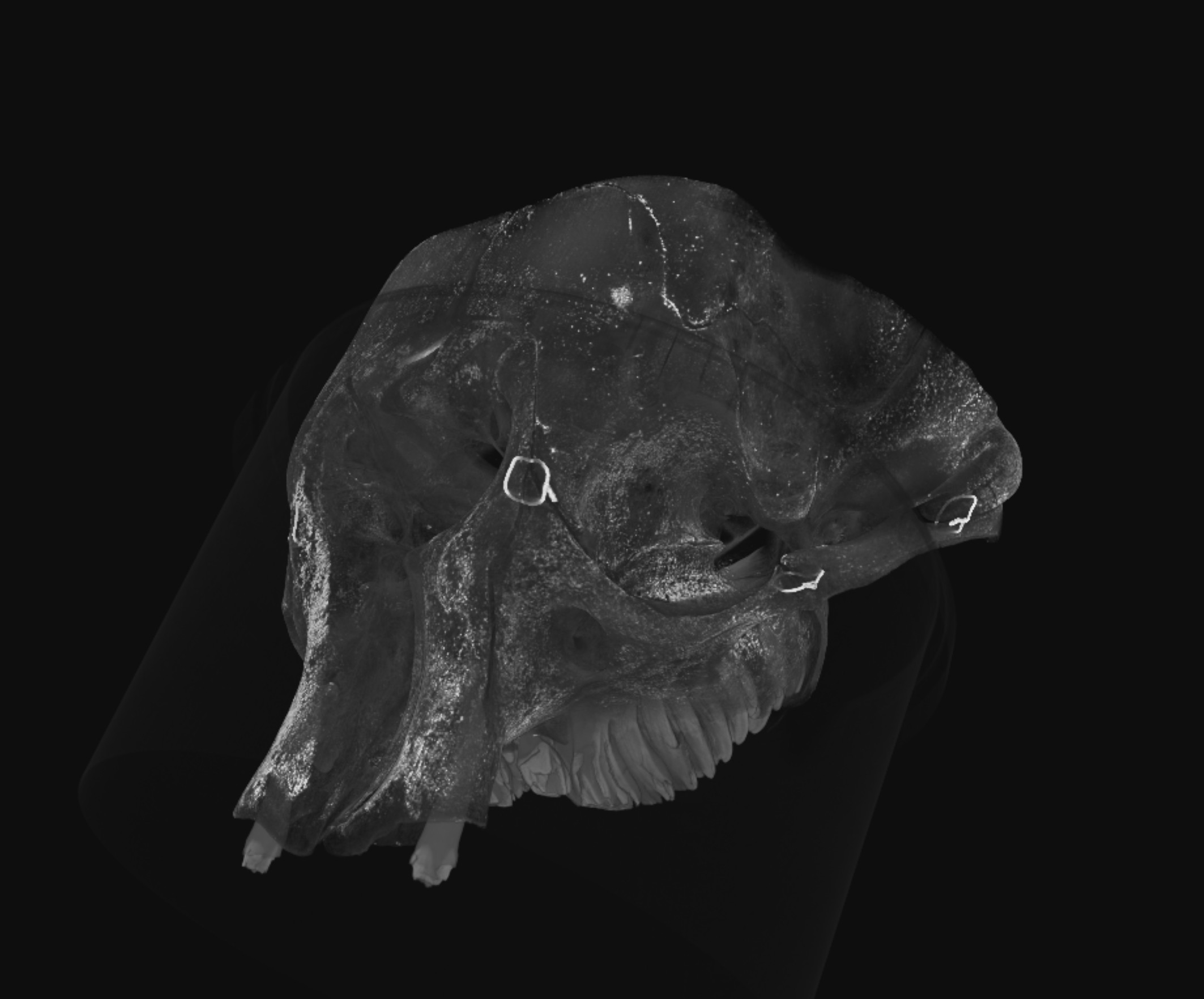
Volume rendering of both parts of the Asian elephant skull aligned.Volume rendering of the microCT scan of the first part ("skull 1") of the Asian elephant skull - this part contains the inner ear.Volume rendering of the microCT scan of the second part ("skull 2") of the Asian elephant skull.

 https://orcid.org/0000-0001-5307-3420